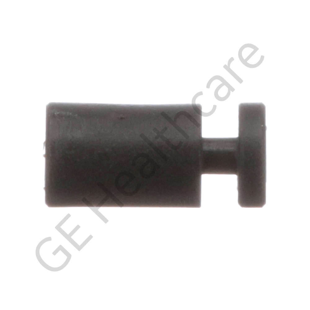

SOCKET
| FB307376 | |
| Ultrasound | |
| GE HealthCare | |
| GE HealthCare | |
Insira o seu número de aprovação e submeta para adicionar item (s) no carrinho.
Please enter approval number
OR
Não sabe o seu número de aprovação? Ligue para 800-437-1171
Enter opt 1 para os primeiros três avisos, e tenha o seu ID do Sistema disponível.
Se você adicionar item (s) no carrinho e enviar seu pedido sem o número de aprovação
, a GE irá contactá-lo antes que o seu pedido
seja confirmado para entrega.
Select your approver's name and submit to add item(s) to your cart
Please Select Approver Name
OR
Don't know your approval number? Call 800-437-1171
Enter opt 1 for the first three prompts, and have your System ID available.
If you add item(s) to cart and submit your order without
selecting an approver, GE will contact you before your order
can be confirmed for shipment.
Visão geral do produto
The Socket Wire Gas Spring in Ultrasound is used in L9 frame kit, Wire assembly, Musashi base kit and other medical equipments as applicable. It is lightweight. They are durable and compact with an excellent dimensional stability. It is cylindrical in shape. The product is made from the material which possess corrosion resistant, impact strength, ductility, easy maintenance, flexibility, high performance, safe and increased lifespan. It is a ROHS compliant and is approved for today’s safety standards. It is aesthetic in appearance. The product does not contain any dirt, dust, visible oils, grease, lint, fibers, rust, paper, and chips, cleaning solutions, machining coolant, foreign materials and other contaminants. The GE product is an innovation and technology, which fits well into versatile customer needs. The product is securely packaged inside a high quality packing box to avoid physical damage during transit and labeled with details about the product, Quality Assurance (QA) seal and shipment details.
Produtos Compatíveis

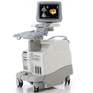
Vivid 7
The Vivid 7/Vivid 7 PRO ultrasound unit is a high performance digital ultrasound imaging system.The system provides image generation in 2D (B) Mode, Color Doppler, Power Doppler (Angio), M-Mode, Color M-Mode, PW and CW Doppler spectra, Tissue Velocity imaging and Contrast applications. The fully digital architecture of the Vivid 7/Vivid 7 PRO unit allows optimal usage of all scanning modes and probe types,throughout the full spectrum of operating frequencies.The Vivid 7 Dimension gives clinicians a better way to convey their findings to other cardiologists, referring physicians,EP physicians and patients. Now, cardiac anatomy,synchronicity and viability can be clearly communicated in imaging formats that are more familiar for your clinical partners, thus easier to understand.
• Real-time 4D imaging – provides more cardiac information to help clinicians better communicate the heart’s structure and function.
• 4D Tissue Synchronization Imaging (TSI) – propels Tissue Velocity Imaging (TVI) to the next level by taking three simultaneous planes – from a single heartbeat at high frame rates – to create a flexible, dynamic 4D model with quantitative measurements to better communicate cardiac dyssynchrony.
• Bull’s-eye report formats and TSI surface mapping –communicate cardiac dyssynchrony in a visual display that should be more familiar to EP physicians.
• Blood Flow Imaging (BFI) – new vascular imaging mode gives clinicians a better understanding and delineation of directional blood flow in vessels.
• Seamless measurement integration – allows you to efficiently calculate ejection fraction and volumes from tri-plane images gathered from the same heartbeat.


